
An ultrasound is one of the most reliable ways to diagnose varicose veins. The large and small veins, branching veins, and interconnected veins in your legs can all be mapped out with ultrasound. Ultrasound is commonly used as a baseline diagnostic tool for varicose veins and as a follow-up following vein, therapy to assess the effectiveness of the procedure. The condition of the valves and the presence of reflux (backflow of blood) or inflammation in a vein can be determined. Blood clots and blocked veins, both partial and total, can be detected by ultrasound which can be treated by a Plano board certified vein and vascular specialist.
In What Ways Does Ultrasound Help?
Using ultrasound, in which high-frequency sound waves are directed at the veins, is a safe, painless, and non-invasive therapy. The frequency shifts as the sound waves interact with the various leg tissues of varying densities. An ultrasound machine can visually represent hidden structures by picking up on the varying sound wave frequencies.
How Will My Doctor Determine If an Ultrasound Is Necessary for Me?
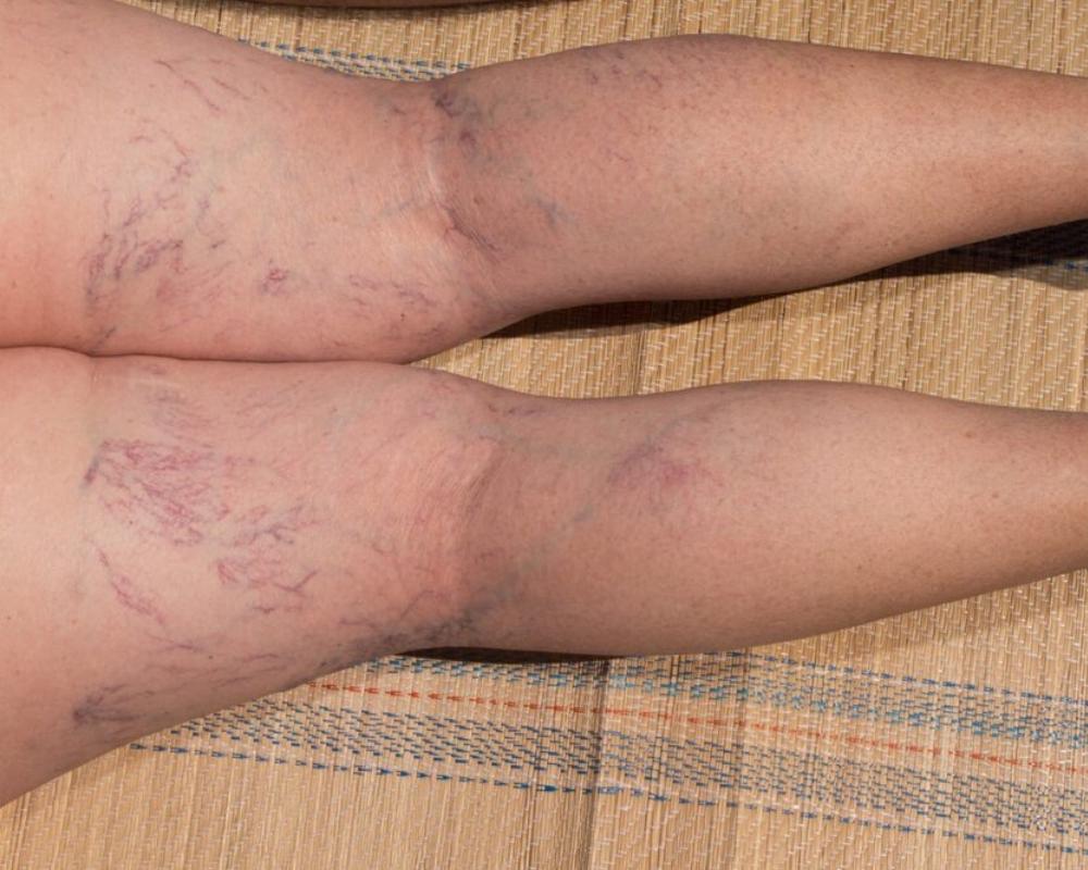
An ultrasound will most likely be necessary if you have symptoms of venous insufficiency. Visible varicose or spider veins; leg or ankle swelling; itchy or painful sensations; or changes in skin tone.
The Ultrasound of My Legs: What Should I Expect?
It usually takes between 30 and 45 minutes to complete a leg ultrasound. A gel that is water-soluble is applied to your skin over the area of interest throughout the process. The tech moves A handheld ultrasound probe around your lower leg. The gel aids in focusing the device’s ultrasonic pulses on the veins.
You will probably be asked to stand up and lie down during the ultrasound. This guarantees the best possible picture quality in three ways:
- Allows for improved ultrasound machine vein access.
- Determines how possible variations in blood flow are affected by changing body posture.
- Measures how well the calf muscle pump is working.
The ultrasound technologist may also apply manual or automated compression to your thigh and leg to evaluate blood flow. In addition, the technician may gently press on the veins to feel for the presence of a clot if a blood clot is suspected in a vein close to the surface or if you are at a greater risk for blood clots.
Veins closer to the skin’s surface are analyzed first, then those in the middle, and finally those further down. An examination of the deep veins is essential if you have edema or skin changes like discoloration, venous eczema, or venous ulcers because it can indicate if a deep vein is compressed and provide evidence of a past blood clot.